Immunofluorescence Protocol For Frozen Sections

Graphic Protocol For The Preparation And Fluorescent Ihc Staining Of Frozen Tissue Sections R D Systems
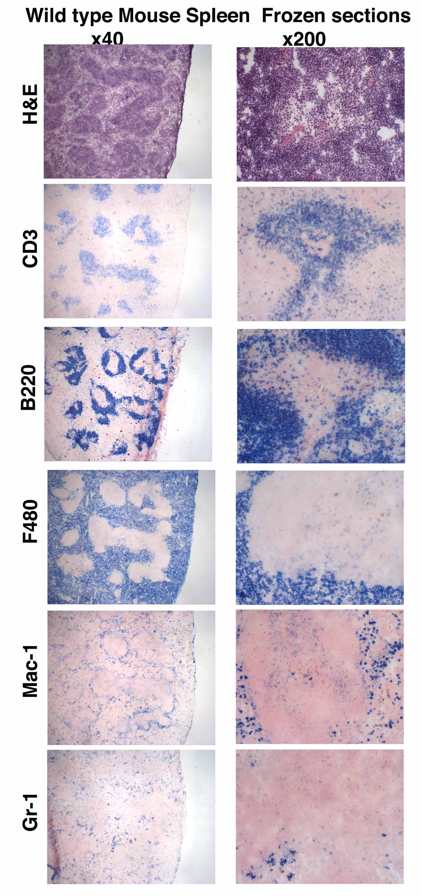
Immunohistochemistry Leinco Technologies
Watch Video Immunofluorescence Tissue Staining Biocompare Streaming Video

Frozen Section Staining For Immunofluorescence If Microscopy Institute For Molecular Bioscience University Of Queensland
Considerations For Immunofluorescence Staining Biotium

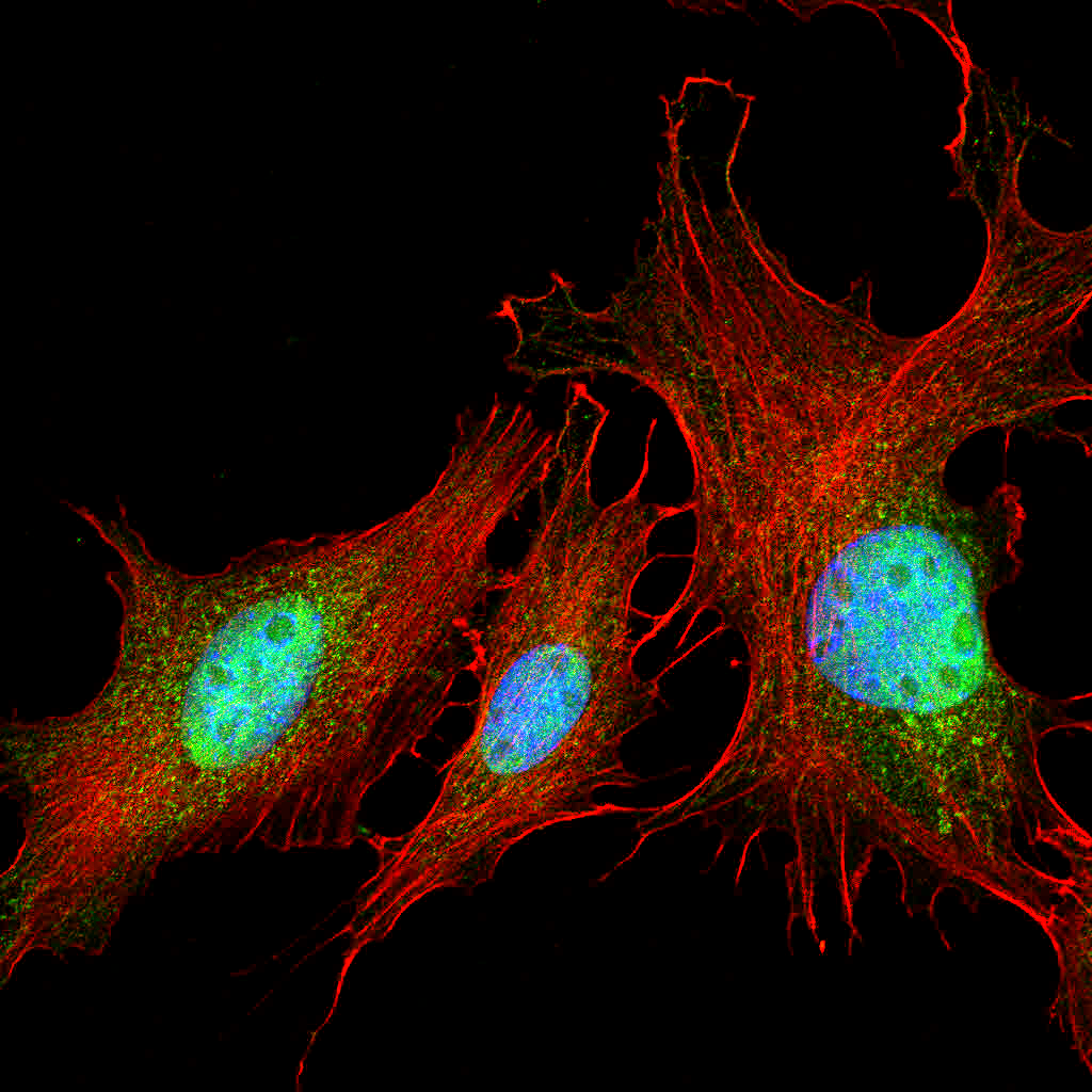
A Manual Multiplex Immunofluorescence Method For Investigating Neurodegenerative Diseases Biorxiv
Cut cryostat sections at 5 10 µm and mount on gelatin coated histological slides.
Immunofluorescence protocol for frozen sections.

Frozen Tissue Slide Preparation And Processing Youtube
Immunofluorescence Troubleshooting Protocol Tips Stressmarq
Confocal Microscopy Staining Brain Cryosections Olympus Life Science
Https Www Jove Com Pdf 59344 Sequential Immunofluorescence Immunohistochemistry On Cryosectioned

What Is The Best Practice Staining On Slides Or Free Floating

Overview Of Immunohistochemistry Thermo Fisher Scientific Ca

2017 Fluomute Ready To Use Reagent To Reduce Autofluorescence In Cells And Tissue Just Incubate Fixed Cells Of Tissue Cells And Tissues Stem Cells Treatment

Cryogenic Tissue Processing And Section Immunofluorescence Of Cerebral Organoids

Immunofluorescence An Overview Sciencedirect Topics

Anti Brdu Antibody Bu1 75 Icr1 Proliferation Marker Ab6326 Abcam

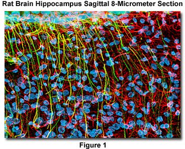
Pin On Moj Toxicology

Anti Gfap Antibody Ab7260 Abcam
Free Floating Or Slide Mounted Immunohistochemistry

Anti Human Nuclear Antigen Antibody 235 1 Ab191181 Abcam

Anti Alpha Smooth Muscle Actin Antibody 1a4 Ko Tested Ab7817 Abcam
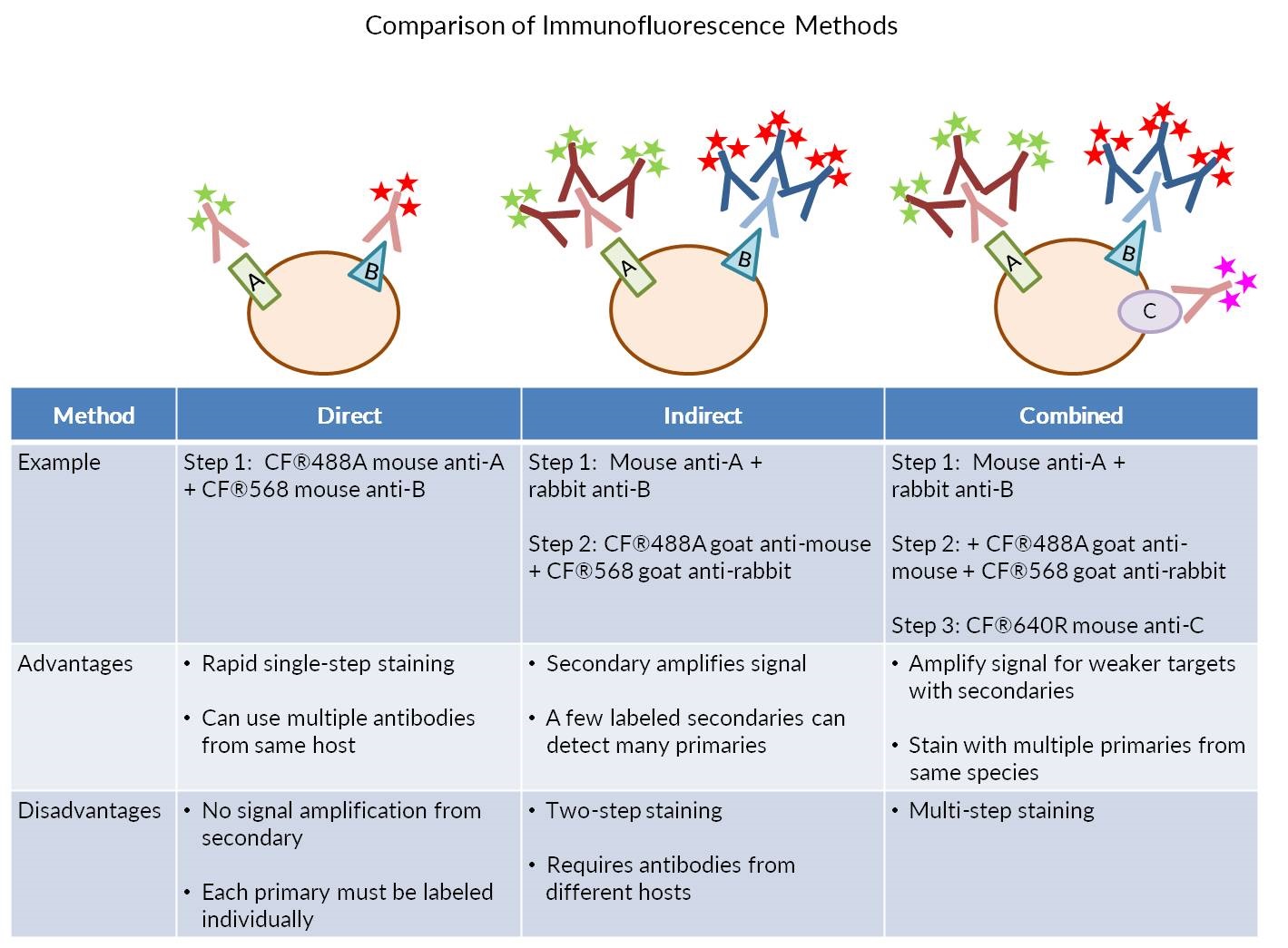
Our Comprehensive Guide To Performing Double Immunofluorescence
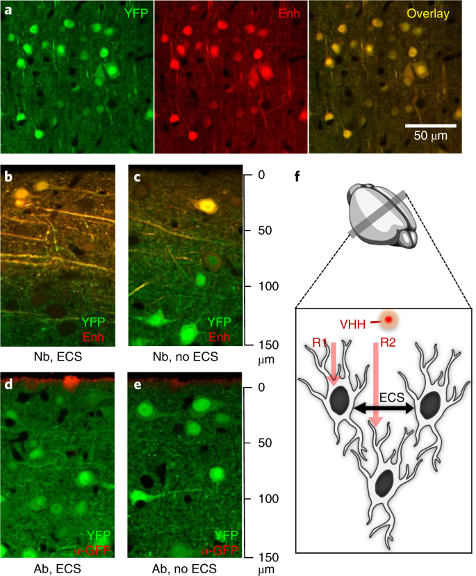
Nanobody Immunostaining For Correlated Light And Electron Microscopy With Preservation Of Ultrastructure Nature Methods

Recombinant Anti Sox9 Antibody Epr14335 78 Ab185966 Abcam
Https Encrypted Tbn0 Gstatic Com Images Q Tbn 3aand9gcsovvalcw Lhxvcxg6jl7eq7c5qqa7spkadlw0ad27m1gwojhyi Usqp Cau
Mouse Phenotyping Ucsd University Of California San Diego

580 Questions With Answers In Immunofluorescence Staining Scientific Method

Anti Myelin Basic Protein Antibody Ab40390 Abcam

Anti Cd68 Antibody Ed1 Ab31630 Abcam

Anti F4 80 Antibody Ci A3 1 Macrophage Marker Ab6640 Abcam
Source : pinterest.com
